Introduction
The Pitt — Episode 4, Stone's block:
"We'll block all the nerves going to the broken ribs. One shot — serratus anterior block down to T9. He'll be awake and pain-free." — Dr. Garcia
"You kids and your crazy regional blocks." — Senior physician
"Yeah, that's what I'm talking about." — Stone, after the procedure
Stone's reaction — from intense pain-driven agitation to near-immediate relief — captures in seconds the clinical impact of the serratus anterior plane block. Dr. Garcia's "crazy regional blocks" comment in The Pitt actually describes a technique that represents the state of the art in chest trauma analgesia: a single ultrasound-guided injection that simultaneously covers 8 to 10 intercostal nerves, without the risks of a thoracic epidural catheter and without the systemic sedation of opioids.
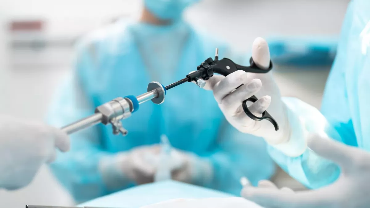
The serratus anterior plane block kit is a relatively simple instrument — block needle, syringe, local anesthetic, ultrasound — but it requires precise anatomical knowledge and careful technique to produce the dramatic result seen in the episode.
What Is the Serratus Anterior Plane Block?
The Serratus Anterior Plane Block (SAPB) is a regional anesthesia technique that deposits local anesthetic in the fascial plane between the serratus anterior muscle and the external intercostal muscles. First described by Blanco et al. in 2013, it rapidly became one of the most widely used regional block techniques in chest trauma, breast surgery, and post-thoracotomy pain management.

The key anatomy: the lateral cutaneous branches of intercostal nerves T2 to T9 emerge from the intercostal space and perforate the serratus anterior muscle to innervate the skin and tissues of the lateral chest wall. By depositing local anesthetic in the plane where these branches emerge, a single injection can block all sensation of the lateral chest wall — from T2 to T9 — which is precisely the region of the fractured ribs in lateral traumas like Stone's.
Block Kit Components
The serratus anterior block requires standard ultrasound-guided regional block equipment:
- Ultrasound with high-frequency linear transducer (6-15MHz): for visualization of chest wall muscle layers — latissimus dorsi, serratus anterior, ribs, and pleura
- Echogenic block needle: 50 to 100mm, 22G short-bevel — adequate length to reach the fascial plane while maintaining ultrasound visualization
- 20-30mL syringe for local anesthetic volume
- Local anesthetic: bupivacaine 0.25% or ropivacaine 0.2-0.375% — 30-40mL for T2-T9 coverage. Bupivacaine lasts 12-18 hours; ropivacaine has a safer cardiovascular profile
- Sterile field, sterile gloves, sterile transducer cover
- Sterile ultrasound gel
Indications
SAPB is indicated for lateral and anterior chest pain:
- Multiple rib fractures: primary indication in trauma — as in Stone's flail chest case
- Blunt chest trauma with chest wall contusion
- Painful chest drainage: analgesia for chest tube or pigtail catheter insertion and maintenance
- Post-thoracotomy — complement to systemic analgesia
- Post-mastectomy and breast surgery
- Thoracic herpes zoster — acute pain relief in post-herpetic neuralgia
Ultrasound Anatomy
Identifying structures on ultrasound follows a logical sequence:
- Position the transducer at the mid-axillary line, at the level of the 4th or 5th ICS, with longitudinal orientation (parallel to ribs)
- Identify the latissimus dorsi muscle — superficial, triangular, moves with respiration
- Identify the serratus anterior muscle — deep to latissimus, with a serrated appearance over the ribs
- Identify the ribs — hyperechoic acoustic shadows with posterior dropout
- Identify the pleura — bright hyperechoic line that slides with respiration (pleural sliding)
- The injection target is the fascial plane between serratus anterior and external intercostals — above the ribs, below the serratus
Block Technique
- Positioning: lateral decubitus with affected side up, or supine with ipsilateral arm abducted to expose the axillary line
- Identify structures on ultrasound as described above
- Antisepsis and sterile field
- Skin local anesthesia: subcutaneous 1% lidocaine at the needle entry point
- In-plane needle insertion with the transducer — lateral-to-medial approach to maintain full needle visualization
- Advance the needle to the plane between serratus anterior and external intercostals — confirmed by visualization of the tip in the correct plane
- Negative aspiration before injecting — exclude intravascular or intrapleural position
- Slow local anesthetic injection: 30-40mL in 5mL increments with periodic aspiration. Observe anesthetic spread between muscle planes on ultrasound — a "separation" appearance between serratus and intercostals
- Needle removal and dressing
- Assess onset: complete sensory block in 10 to 20 minutes — test with pinching of T2-T9 dermatomes
Advantages Over Alternative Techniques
SAPB has important advantages over traditional alternatives for rib fracture analgesia:
- vs. Thoracic epidural catheter: lower hypotension risk, no anticoagulation contraindication, technically simpler, can be performed in supine position
- vs. Paravertebral block: lower pneumothorax risk, shallower insertion depth, easier ultrasound visualization of structures
- vs. Multiple intercostal blocks: one injection covers 8 to 10 dermatomes vs. one injection per fractured rib — dramatic reduction in procedures, time, and total anesthetic dose
- vs. Systemic opioids: comparable analgesia without sedation, respiratory depression, or constipation — extremely relevant in chest trauma where ventilation is already compromised
Prognosis & Complications
SAPB has an excellent safety profile when ultrasound-guided. Studies show 40-70% reduction in opioid consumption in the first 24 hours after the block for rib fractures.
Possible complications:
- Pneumothorax: rare with ultrasound guidance — real-time pleural visualization prevents pleural penetration
- Local anesthetic systemic toxicity (LAST): due to the high dose (30-40mL) — fractional injection with periodic aspiration is mandatory; have intralipid available
- Hematoma: in anticoagulated patients — assess hemostasis before the procedure
- Inadequate block: insufficient anesthetic spread — may require needle repositioning
- Limited duration: bupivacaine lasts 12-18 hours — may require repetition or continuous catheter for prolonged analgesia

Frequently Asked Questions
Can any emergency physician perform SAPB?
SAPB is considered an intermediate-difficulty block in regional anesthesia. It requires specific training in ultrasound-guided regional anesthesia. In many trauma centers, it is performed by anesthesiologists or pain physicians with advanced POCUS training. The growing trend is to include SAPB in the advanced emergency medicine curriculum, given its impact on chest trauma management.
Why does Dr. Garcia mention blocking "down to T9"?
SAPB coverage depends on injection volume and position. Volumes of 30-40mL in the serratus anterior plane typically cover T2 to T9 — the entire lateral chest wall. Stone had multiple rib fractures throughout the lateral extent of the left hemithorax, so T2-T9 coverage was exactly what was needed to treat all fractured ribs with a single injection.
Can SAPB be repeated?
Yes. The block can be repeated when the anesthetic effect begins to wane (typically after 12-18h with bupivacaine). Alternatively, a continuous catheter can be left in the serratus plane for continuous local anesthetic infusion — a technique used in ICU patients with multiple rib fractures requiring prolonged analgesia.
Can the block be performed in anticoagulated patients?
SAPB is considered a low hematoma risk block compared to epidural or paravertebral — the fascial plane between serratus and intercostals is relatively superficial and compressible. ASRA regional anesthesia guidelines classify SAPB as a superficial block, where anticoagulation is not an absolute contraindication. However, clinical context and bleeding risk should be assessed individually.
Conclusion
The serratus anterior block in The Pitt is presented as a "crazy regional block" — but Stone's reaction proves the opposite: it is precision analgesia, image-guided, with broad coverage, minimal risks, and maximum comfort for a patient who would otherwise need escalating opioid doses or an invasive epidural catheter. It is the future of chest trauma analgesia — and already the present in advanced trauma centers.
See also: Flail Chest Scenario: From Serratus Block to Pneumothorax, Flail Chest Pathophysiology, and Point-of-Care Ultrasound in Emergency.
This content is for educational purposes only and does not substitute professional medical evaluation, diagnosis, or treatment. In case of emergency, call 911 immediately.